
The world’s most poisonous mushroom, Amanita phalloides, is growing in BC
Issue: BCMJ,
vol. 61 , No. 1 , January February 2019 ,
Pages 20-24 Clinical Articles
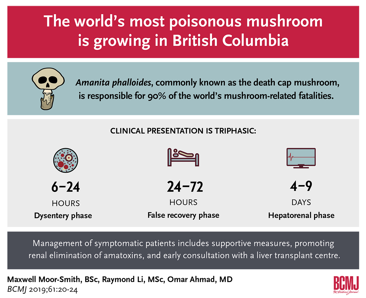
ABSTRACT: Amatoxins in Amanita phalloides, commonly known as the death cap mushroom, are responsible for 90% of the world’s mushroom-related fatalities. The most deadly amatoxin for humans is α-amanitin, a bicyclic octapeptide that irreversibly binds RNA polymerase II, thus preventing protein synthesis and causing cell death. Three recent poisoning cases in British Columbia show how the death cap can be easily mistaken for edible mushrooms such as the puffball and the paddy straw mushroom. Since being introduced from Europe to the west and mid-Atlantic coasts of North America, A. phalloides has spread to south coastal BC, and has the potential to spread to vast areas of the continent. Following ingestion of A. phalloides, there is a latency period (6 hours) followed by intoxication, classically described as triphasic: a dysentery phase (6 to 24 hours), a false recovery phase (24 to 72 hours), and a hepatorenal phase (4 to 9 days) consisting of multisystem organ failure, seizures, coma, and death.
Treatment is based on decontamination and liver transplantation if acute liver failure occurs. Management of the symptomatic patient consists of providing supportive care, promoting renal elimination of amatoxins, interrupting enterohepatic recirculation of amatoxins, and administering proposed antidotes. Although no established antidote for A. phalloides has been identified, N-acetylcysteine and silibinin have shown some benefit in a retrospective survival analysis. With the expanded range of A. phalloides in BC, physicians should be alert to the possibility of amatoxin poisoning and include it in the differential diagnosis of a patient presenting with gastroenteritis or hepatotoxicity and a history of ingesting foraged mushrooms.
The expanded range of death cap mushrooms—previously found on the roots of imported European trees but now found in association with native Garry oaks—puts amateur foragers at risk, and recognition of amatoxin poisoning is essential to preventing future fatalities.
Amanita phalloides, known commonly as the death cap mushroom, causes life-threatening hepatorenal dysfunction when ingested. Considered the most poisonous mushroom in the world, A. phalloides contains amatoxins, a group of bicyclic octapeptides that are responsible for 90% of global mushroom-related fatalities. One cap of A. phalloides is sufficient to cause death in an adult.[1-3]
The death cap was first introduced to British Columbia on the roots of imported European trees and has since spread to North American oak trees.[4,5] Death caps are now found increasingly in urban settings. In 2017 the Canadian Forest Service and Oak Bay parks department reported that death caps in the Victoria area sprouted earlier and in greater numbers than in previous years.[6] The spread of this invasive species has led to cases of morbidity and mortality from ingestion of the mushroom and an ongoing risk of misidentification. Health care providers need to be aware of this risk, as prompt recognition and appropriate management are critical for positive patient outcomes.
Distribution
A. phalloides is not native to North America. First identified in Europe, the species has now traveled to Australia, Asia, Southern Africa, and the Americas on the roots of imported trees.[5] The first confirmed collection of A. phalloides in North America was in northern California at the Hotel Del Monte in 1935, a location famous for its exotic and unusual gardens.[5] Since then, the death cap has been introduced to multiple sites in the Pacific Northwest.
A. phalloides specimens were first collected in BC in 1997 from under European chestnut trees at Lake Errock in the upper Fraser Valley. The first identification of A. phalloides in Victoria was in 1998 from under a large European beech tree on the landscaped grounds of Government House, the residence of BC’s lieutenant governor. A. phalloides was detected in Vancouver in 2008 under European hornbeam trees that had been planted by the city in the 1960s.[4] Since these first specimens were collected, there have been numerous reports of A. phalloides found in Vancouver and the Fraser Valley, on Southern Vancouver Island, and on the Gulf Islands.
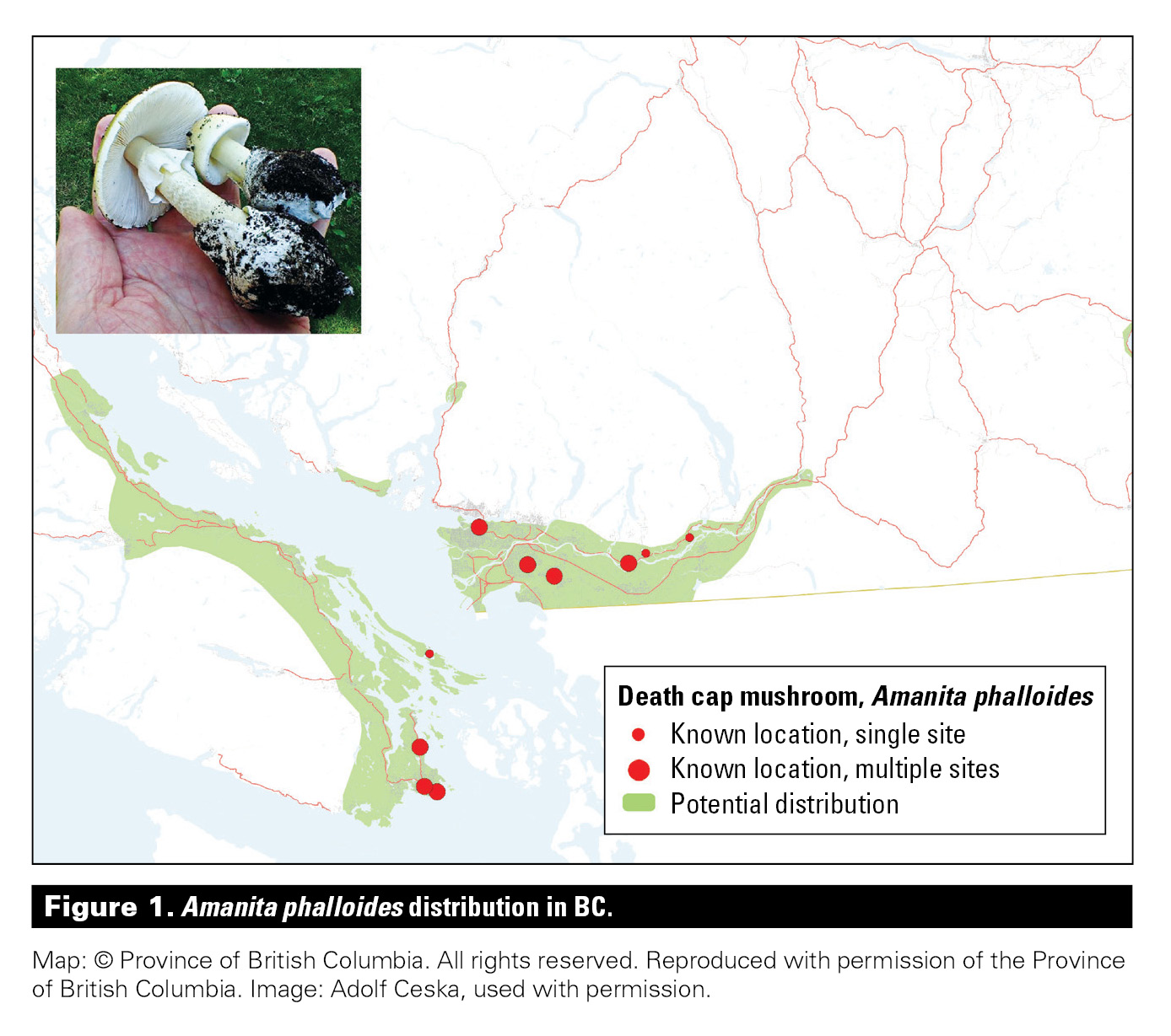
As an ectomycorrhizal fungus, A. phalloides forms an obligate symbiotic relationship with the roots of trees, which have been mostly nonnative, broad-leaf trees.[4,5] This association may have limited the mushroom’s spread so far. However, in Victoria A. phalloides has now been found in association with native BC Garry oak trees, which may allow the mushroom to expand its range even further.[4] Pringle and colleagues estimate the speed of A. phalloides spread among native California coastal oak at 5 km per year on average (range 3 to 9 km/year).[5] Additionally, predictive climate suitability mapping shows that most of south coastal BC is appropriate habitat for A. phalloides[5] (Figure 1).
Mushroom foragers and health care providers should be aware of the expanded range for this highly toxic mushroom in order to prevent fatalities from the death cap in the future.[7]
Identification and misidentification
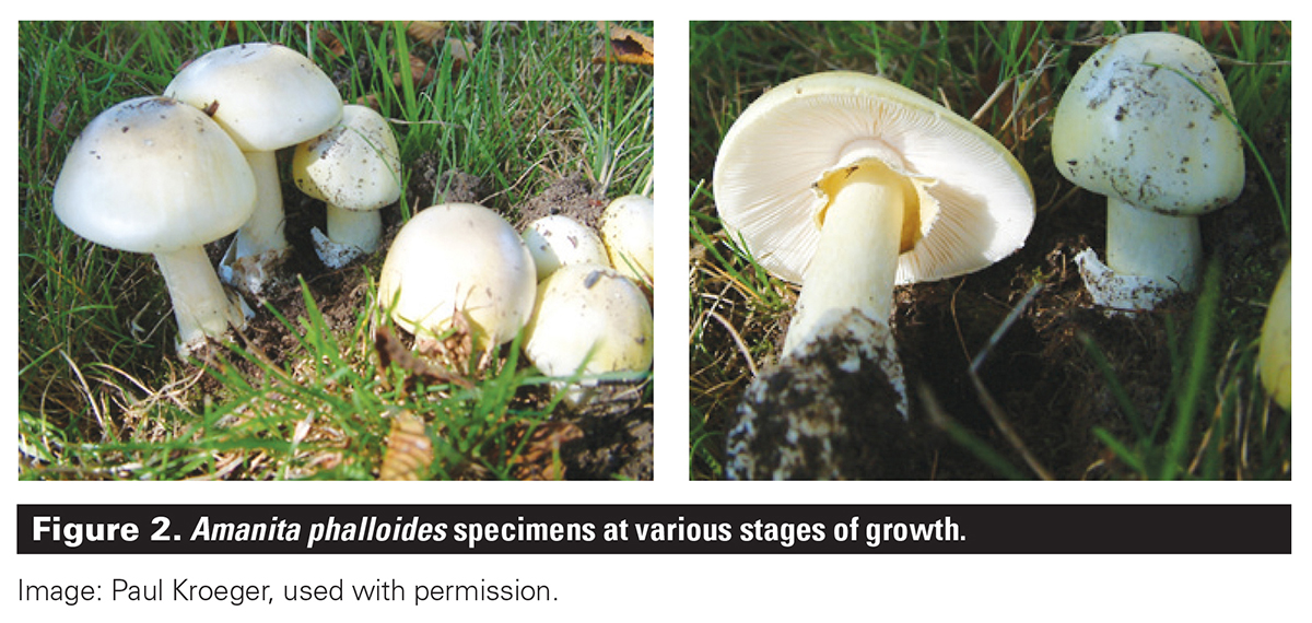
A. phalloides typically grows from June to November in BC and looks different depending on its stage of maturity. The immature mushroom may be totally encased in a “universal veil,” giving it the egg-like appearance of a puffball mushroom if not cut in half to reveal the growing mushroom inside. As the mushroom matures, the universal veil ruptures and remains in the ground as a membranous sac (volva). The cap may grow up to 16 cm in diameter, becoming broad and slightly convex. The cap can be white or have a green or yellow hue, and is often darker in the centre. A. phalloides specimens typically feature white gills and spores, a ring of tissue (annulus) near the top of the stalk, and a volva at the base[8] (Figure 2).
Death caps can easily be misidentified as an edible variety of mushroom, as seen in three cases of amateur foragers who mistook the death cap for other species. In 2003 a 43-year-old man in Victoria consumed an immature death cap he thought was a puffball mushroom. In 2008 a 63-year-old woman in Vancouver consumed a mature death cap she assumed was a paddy straw mushroom, a common variety in Asia but one not native to North America. Both patients recovered following hospitalization. In 2016 a 3-year-old boy died after consuming a death cap foraged from a residential street in Victoria.[4]
Toxicity
Three classes of toxins are present in A. phalloides: amatoxins, phallotoxins, and virotoxins. Of the three, amatoxins exert the greatest effect, and the most toxic for humans is α-amanitin.[1,2,8] Like all the major amanitins (α, β, γ), α-amanitin is a bicyclic octapeptide that irreversibly binds RNA polymerase II, thus preventing protein synthesis and causing cell death. None of the amatoxins are destroyed by cooking, drying, or freezing. The presence of organic anion transporting polypeptide 1B3 (OATP1B3) in hepatic sinusoidal membranes results in the active transport of toxin into hepatocytes, causing massive centrilobular necrosis and vascular degeneration.[2,9] Amatoxins are primarily eliminated by renal excretion, with a portion undergoing biliary excretion and enterohepatic recirculation.[9] The kidneys may also be affected and show signs of acute tubular necrosis and hyaline casts in the tubules.[2]
Triphasic clinical presentation
Symptoms following the ingestion of nonfatal mushroom species generally occur within 6 hours. In contrast, symptoms of A. phalloides poisoning arise 6 to 24 hours after ingestion. Symptoms occurring within 6 hours of ingestion do not exclude the possibility of A. phalloides ingestion, however, as multiple species of foraged toxic mushrooms are often ingested together.[3] After this initial latency period, there are three phases in the clinical presentation of A. phalloides poisoning.
Most patients present in the first (dysentery) phase, which is characterized by abdominal pain, vomiting, and severe, cholera-like diarrhea that may contain blood and mucus, and often results in profound dehydration.[2,10] The second (false recovery) phase occurs 24 to 72 hours after ingestion with the patient demonstrating symptomatic improvement despite clinical signs and biochemical markers of liver damage progressing, peaking at 60 to 72 hours after ingestion.[11] The third (hepatorenal) phase occurs 4 to 9 days after ingestion and is characterized by acute liver and multisystem organ failure that can lead to convulsions, hemorrhage, coma, and death.[2,3]
Diagnosis and management
Amatoxin mushroom poisoning can be fatal. The best prognosis results from prompt recognition and appropriate management. The foundation of diagnosis is an accurate history and recognition of the toxidrome. Specimens or photographs of the mushroom consumed can help confirm the diagnosis, but often samples are partially decomposed or do not represent the ingested species. Assays to detect amatoxins are not available locally.[3]
For asymptomatic patients, gastrointestinal decontamination with activated charcoal should be considered, even if the patient presents several hours after ingestion.[9] For symptomatic patients, management should include providing supportive care, promoting renal elimination of amatoxins, interrupting enterohepatic recirculation of amatoxins, and administering proposed antidotes (Table).[12-14] In addition, early consultation with the local poison control centre and a liver transplant centre is advised.
No established antidote for amatoxin poisoning has been identified. Several have been proposed, but their efficacy is not proven. A meta-analysis of 2108 hospitalized patients with amatoxin poisoning found therapies with silibinin or the hepatoprotectant N-acetylcysteine (NAC) were the most effective in a retrospective survival analysis.[2]
Silibinin (extract of milk thistle) inhibits OATP1B3 and prevents uptake of amatoxins into hepatocytes.[9] While intravenous silibinin (Legalon SIL) has been used in Europe for many years, in Canada it is only available through the Special Access Programme. Oral silibinin is available at health food stores, but it may be inactivated when given with activated charcoal. High-dose penicillin G and cyclosporine are other potential antidotes as they inhibit OATP1B3 and thus can prevent transport of amatoxin into hepatocytes. Penicillin has been one of the most commonly used therapies, despite showing limited benefit in a retrospective survival analysis.[2] Although the risks associated with high-dose penicillin (hypernatremia, seizures) are more significant than other proposed therapies, penicillin use can be considered for hepatoprotection if IV silibinin is not an option.[9]
N-acetylcysteine is thought to limit hepatic damage through its free-radical or oxygen scavenging capabilities. Due to the benign side-effect profile of NAC, the benefits of its use are thought to outweigh any risks.[7,13]
Once acute liver failure occurs, liver transplantation is the only definitive treatment.[1,3,10,14]
Summary
The physician’s role in preventing fatalities from A. phalloides ingestion lies in prompt recognition of amatoxin poisoning in a patient. Clinicians should be particularly alert if the patient reports consuming foraged puffball mushrooms or paddy straw mushrooms, as both of these mushrooms are known to resemble the death cap. With the expanded range of A. phalloides in BC, physicians should include amatoxin toxicity in the differential diagnosis of a patient presenting with gastroenteritis or hepatotoxicity and a history of ingesting foraged mushrooms. Management of the symptomatic patient involves providing supportive measures, promoting renal elimination of amatoxins, interrupting enterohepatic recirculation of amatoxins, administering proposed antidotes, and consulting with the local poison control centre and a liver transplant centre.
Acknowledgments
The authors would like to thank Dr Daniel Ovakim for his assistance in developing this article. Dr Ovakim is affiliated with Critical Care Medicine at Island Health, and Medical Toxicology at the BC Drug and Poison Information Centre.
Competing interests
None declared.
This article has been peer reviewed.
References
1. Garcia J, Costa VM, Carvalho ATP, et al. A breakthrough on Amanita phalloides poisoning: An effective antidotal effect by polymyxin B. Arch Toxicol 2015;89:2305-2323.
2. Enjalbert F, Rapior S, Nouguier-Soulé J, et al. Treatment of amatoxin poisoning: 20-year retrospective analysis. J Toxicol Clin Toxicol 2002;40:715-757.
3. Berger KJ, Guss DA. Mycotoxins revisited: Part I. J Emerg Med 2005;28:53-62.
4. Berch SM, Kroeger P, Finston T. The death cap mushroom (Amanita phalloides) moves to a native tree in Victoria, British Columbia. Botany 2017;95:435-440.
5. Pringle A, Adams RI, Cross HB, Bruns TD. The ectomycorrhizal fungus Amanita phalloides was introduced and is expanding its range on the west coast of North America. Mol Ecol 2009;18:817-833.
6. Brend Y. Death cap mushrooms boom 1 year after child’s death in Victoria. CBC News. 11 October 2017. Accessed 20 November 2018. www.cbc.ca/news/canada/british-columbia/death-cap-mushroom-boom-deadly-fungi-victoria-oak-bay-garry-oak-1.4349883.
7. Ward J, Do KK, Brush E, Salhanick SD. Amatoxin poisoning: Case reports and review of current therapies. J Emerg Med 2013;44:116-121.
8. French LK, Hendrickson RG, Horowitz BZ. Amanita phalloides poisoning. Clin Toxicol (Phila) 2011;49:128-129.
9. Zilker T, Faulstich H. Cyclopeptide-containing mushrooms: The deadly Amanitas. In: Brent J, Burkhart K, Dargan P, et al. (eds). Critical care toxicology. 2nd ed. New York: Springer; 2017. p. 2129-2148.
10. Diaz JH. Syndromic diagnosis and management of confirmed mushroom poisonings. Crit Care Med 2005;33:427-436.
11. Giannini L, Vannacci A, Missanelli A, et al. Amatoxin poisoning: A 15-year retrospective analysis and follow-up evaluation of 105 patients. Clin Toxicol (Phila) 2007;45:539-542.
12. Kieslichova E, Frankova S, Protus M, et al. Acute liver failure due to Amanita phalloides poisoning: Therapeutic approach and outcome. Transplant Proc 2018;50:192-197.
13. Stein CM, Wu PE, Scott JA, Weinerman AS. Fulminant hepatic failure following ingestion of wild mushrooms. CMAJ 2015;187:822-824.
14. Vo KT, Montgomery M, Mitchell ST, et. al. Amanita phalloides mushroom poisonings—Northern California December 2016. MMWR Morb Mortal Wkly Rep 2017;66:549-553.
Mr Moor-Smith is a medical student (class of 2020) in the University of British Columbia Island Medical Program. Mr Li is a drug and poison information pharmacist at the BC Drug and Poison Information Centre. Dr Ahmad is a physician with Island Health and head of Critical Care and Emergency Medicine. He is also a clinical associate professor in the Department of Emergency Medicine at the University of British Columbia.


Very helpful