Proposal to improve rectal cancer outcomes in BC
Issue: BCMJ,
vol. 45 , No. 7 , September 2003 ,
Pages 330-335 Clinical Articles
We have identified a problem with high local recurrence rates for rectal cancer in BC in 1996. Factors contributing to poor results are identified including suboptimal use of staging investigations, adjuvant therapy, precise surgical technique (total mesorectal excision), and total mesorectal excision pathology reporting. We have begun education programs for surgeons and pathologists. BC Cancer Agency Surgical Oncology Network is supporting education and prospective data collection in order to improve our outcomes.
Colorectal cancer is a major cause of death in BC. With 800 new cases of rectal cancer spread across the 95 million hectares of the province of British Columbia, changing clinical practice will be a considerable challenge. The good news is that we have a vision of improving outcomes using a coordinated approach to implement practice standards and changes in clinical practice.
Consider the case of a 59-year-old male who presents with a 3-month history of rectal bleeding and increasing constipation. On examination, a palpable rectal tumor of 4 cm diameter is found. The tumor is located on the posterior wall of the rectum 5 cm from the anal verge. By palpation the tumor is mobile but invades the full thickness of the bowel wall into the perirectal fat. Endoscopy and biopsy are performed to confirm the diagnosis of adenocarcinoma. The patient is brought urgently to the operating room for abdominoperineal resection. The surgery and immediate postoperative course are uncomplicated. The pathology is returned reporting clear proximal and distal margins. The depth of invasion is full thickness bowel wall into the perirectal fat. Four of nine lymph nodes contain metastatic tumor. On this basis, the patient is treated with postoperative adjuvant radiation and chemotherapy. A problem arises 18 months later when a rising CEA level prompts investigation. A CT scan shows recurrence of pelvic cancer. The following questions need to be answered:
• Did the patient receive optimal management?
• Was preoperative radiology imaging—such as CT or MR and endorectal ultrasound—considered in order to assess depth of invasion, presence of metastases, and closeness of the tumor to the pelvic sidewall margins?
• Was adjuvant preoperative radiation and chemotherapy considered?
• Was total mesorectal excision (TME) performed? What about radial margin pathology?
Intact mesorectal fascial envelope and a radial margin of more than 2 mm are associated with local recurrence of less than 10%. With TME as the surgical technique, preoperative short-course radiation has been shown to decrease local recurrence to 4% to 5% in prospective national Dutch and Swedish trials.[1,2]
From our review of clinical outcomes of rectal cancer management in BC in 1996,[3] we have identified a problem of high local recurrence rates (Table 1). Local recurrence rates at 4 years in BC are much higher than recurrence rates published for the recent large national Dutch trial of preoperative radiation and TME surgery.[1] We have analyzed potential causes for this high local recurrence rate and have reviewed similar published reports from Sweden,[2] the Netherlands,[1] and Norway[4] in order to design strategies aimed to improve local recurrence rates.
As explained in the previous article, “Practice patterns and appropriateness of care for rectal cancer management in British Columbia,” we did not routinely use staging investigations for rectal cancer assessment. For example, CT was used in about 23% of cases. In association with this lack of radiologic assessment of tumor stage, most adjuvant therapy was given postoperatively. Further, adjuvant therapy was not given in a significant proportion of cases. For example, 30% of patients with stage 3 rectal cancer did not receive adjuvant radiation and 20% of patients with stage 3 cancer did not receive adjuvant chemotherapy.
In a previous review of rectal cancer management, we identified that distance of the rectal cancer from the anus was a factor significantly influencing local recurrence.[3] Tumors more than 10 cm from the anus had a local recurrence rate of less than 10%. In contrast, tumors 10 cm or less from the anus had a local recurrence rate of about 30%. We hypothesized that these low rectal cancers had higher local recurrence on the basis of higher residual tumor status due to increased technical surgical difficulty to achieve a negative radial resection margin in the small confines of the narrow distal pelvis. In support of this hypothesis, the Dutch have reported that their incomplete TME specimen rates increased with decreasing distance from the anus and that an incomplete TME specimen was associated with increased local recurrence (Table 2).[5] However, when we attempted to assess the presence of residual disease at radial margins in this series we found that neither operative nor pathology reports consistently assessed macroscopic or microscopic radial margins. In addition, the technique of TME for the rectal cancer resective surgery was not consistently used in operative or pathology reports. Therefore, it is likely that TME was not performed as a standard preferred rectal cancer resection technique and our pathologists were not trained to report on the intactness of the TME specimen or on radial margins.
Swedish, Dutch, and Norwegian physicians have successfully introduced changes in rectal cancer management as national policies.[1,2,4] They have adopted TME as the standard operation for rectal cancer in their countries. They employed Dr Bill Heald (Basingstoke, UK), Dr Brendan Moran (Basingstoke, UK) and Dr Phil Quirke (Leeds, UK) to teach TME techniques to their surgeons and pathologists.[2,6-8] Whereas local recurrence rates for rectal cancer in these countries were about 30% to 40% before the change to TME surgery, after TME training local recurrence rates fell to less than 10%. Therefore, if we wish to improve local recurrence rates in BC, we too should change our rectal cancer management protocols to include preoperative radiation and TME surgical and pathology techniques.
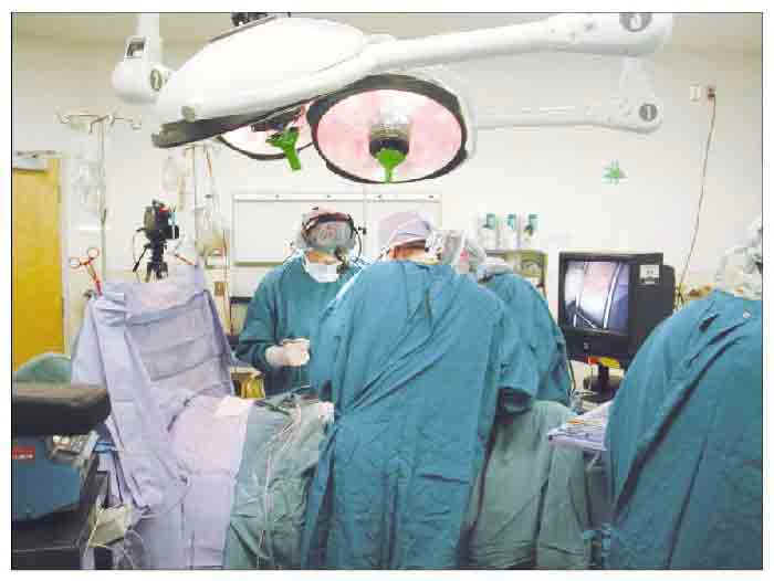
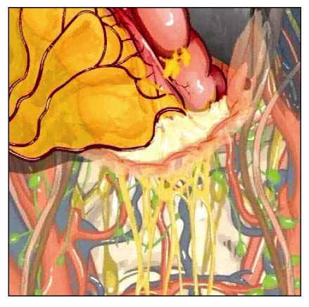
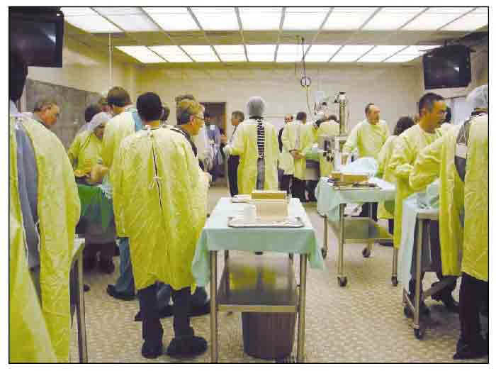
In our consideration of strategies to improve our outcomes for rectal cancer, we have published guidelines for rectal cancer management in this theme issue of the BC Medical Journal (2003;45[6]:264) and on the BC Cancer Agency web site (www.bccancer.bc.ca). We organized a well-attended, technically oriented TME and interdisciplinary course for rectal cancer management in Vancouver in November 2002. The first day of the conference was designed as a technically oriented course for 60 general surgeons to review pelvic anatomy, the technique of TME, and pathology of the TME specimen. Brendan Moran, a consultant surgeon who works with Bill Heald in Basingstoke, UK, presented the rationale for TME (that is, TME is the preferred surgical technique for rectal cancer excision as it results in the lowest reported recurrence rates). Carolyn Compton, a pathologist from McGill, presented convincing data associating an intact mesorectal fascial envelope from TME with the lowest reported local recurrence rates. Terry Phang reviewed pelvic anatomy (see the figure on page 315), and TME surgical techniques with cadaver photos and video and with a live OR broadcast of a TME operation (Figure 1). The case was a 63-year-old male diagnosed with a rectal cancer on the anterior wall within 2 cm of the anal sphincter. Preoperative CT and endorectal ultrasound assessed the tumor as stage T3N1 occupying 75% of the rectal circumference. Because of clinical anterior fixation of the tumor and in an attempt to downstage the tumor in order to preserve the anal sphincter, the patient underwent long-course preoperative radiation and chemotherapy. The TME operation was performed by Drs Phang and Moran with live broadcast of the surgery and interactive discussion with the course participants led by John Macfarlane. The TME surgery demonstrated pelvic nerve preservation, a “glitzy, Hollywood TME specimen” with intact circumferential TME mesorectal fascial envelope, anal sphincter preservation, and reconstruction by a colonic pouch anal anastomosis. The final pathology showed a T2N0R0 cancer with distal resection margin of 1 cm and radial margin of 3 mm. There is no known residual cancer in this patient. Karim Qayumi presented an animated video by artist Dana Smith of TME surgery (Figure 2). Course participants went to the UBC anatomy lab for hands-on cadaver pelvic dissection with an emphasis on identifying pelvic autonomic nerves (Figure 3). In small group sessions, 12 surgeon-instructors assisted course participants with the dissection and with discussion of TME techniques and difficult rectal cancer cases. These instructors included international, national, and local surgeons with extra training in colorectal surgery or surgical oncology: Drs Brendan Moran (UK), Cornelis van de Velde (the Netherlands), Mark Kimmins (Seattle), Jean Couture (Sherbrooke), Marko Simunovic (Hamilton), Don Buie (Calgary), Will Orrom (Victoria), Geoff Tse (Surrey), James Okamura (Burnaby), Rona Cheifetz (VGH), Greg McGregor (VGH), John MacFarlane (St. Paul’s), and Terry Phang (St. Paul’s). An added feature of this technical surgical course was a pre- and post-test designed by Drs Rona Cheifetz and Terry Phang.
The second day of the TME course extended discussion to interdisciplinary management of rectal cancer with 145 attendees, including general surgeons, pathologists, radiologists, radiation and medical oncologists, and UBC residents. Dr Cornelis van de Velde, principal coordinator of the Dutch trial, reviewed data proving effectiveness of preoperative short-course radiation and TME for reducing local recurrence. Dr Phil Quirke, the originator and principal advocate of TME pathology from Leeds, UK, demonstrated processing of the TME specimen to pathologists attending the course. Dr Terry Phang presented results of an audit of rectal cancer pathology reporting for 1996 and 2000. Dr Peter Zetler, a pathologist from St. Paul’s Hospital, proposed a reporting template for BC pathologists. Dr Jacqueline Brown, a GI radiologist from St. Paul’s Hospital, reviewed use of CT, MR, and endorectal ultrasound for staging rectal cancer and assessing need for considering preoperative radiation. Drs John Hay and Amil Shah from the BC Cancer Agency reviewed recommendations for preoperative adjuvant radiation and chemotherapy. Dr Don Buie, a colorectal surgeon from Calgary, spoke on approaches to locally recurrent rectal cancer. Various case presentations of rectal cancer—including the malignant polyp, early rectal cancer, typical rectal cancer, and liver metastases—were discussed in small group sessions led by 12 surgeon-instructors. The concluding session of the course addressed organization to improve our outcomes for rectal cancer. Dr Couture spoke on our Canadian NCIC rectal cancer trial group’s effort to enroll patients into the MRC (UK) study of TME and adjuvant therapy. Dr Simunovic reported his preliminary data of TME training in Ontario, which showed that some surgeons were resistant to learning TME. Dr Phang concluded the meeting with a synthesis of the presented information and an audience discussion of strategies and barriers to improving outcomes for rectal cancer management in BC.
The BC Provincial Surgical Oncology Council and Network has established a colorectal tumor site group to further our mission of implementing changes aimed at improving rectal cancer outcomes in BC. We have started a dialogue with surgeons representing high-volume hospitals in BC (performing 10 or more rectal cancer resections in 1996). These hospitals are as follows:
• Vancouver Coastal region—Vancouver Hospital, St. Paul’s Hospital, Mount St Joseph Hospital, Richmond Hospital, Lions Gate Hospital.
• Fraser region—Burnaby Hospital, Royal Columbian Hospital, Surrey Hospital, Abbotsford Hospital.
• Interior region—Royal Inland Hospital (Kamloops), Kelowna General Hospital.
• Northern region—Prince George Hospital.
• Vancouver Island region—Victoria General Hospital, Royal Jubilee Hospital, Nanaimo General Hospital.
This representation is for the purpose of initial discussion. It will capture about 80% of rectal cancer patients in BC. It is not meant to be exclusive. Minutes of this dialogue will be sent to BC Surgical Society general surgeons.
In order to replicate changes used to improve rectal cancer outcomes in Sweden, the Netherlands, and Norway, we are recommending that general surgeons have TME training by attending a technically oriented TME course and by having a proctor assist at a rectal cancer operation. Representative surgeons from high-volume rectal cancer hospitals have been asked to assess their own hospital divisions of general surgery to determine which surgeons have attended a technical TME course and the need for holding further TME training courses. We have discussed the need to establish funding for a system of proctors to travel to attend rectal cancer surgery throughout BC. Representative surgeons have been asked to communicate with their hospital pathologists and radiologists to make a commitment to TME pathology and preoperative radiologic assessment using CT, MR, and endorectal ultrasound. Where access to these radiologic scans is difficult, we must devise a plan to improve the situation. Representatives from BC Cancer Agency radiation and medical oncology have the task of advocating and implementing preoperative adjuvant therapy for rectal cancer patients in all its BC branches—Vancouver, Surrey, Kelowna, and Victoria.
Pathologist leaders have accepted recommendations to revise pathology reporting for the TME specimen. Representative pathologists from Vancouver Hospital, St. Paul’s Hospital, Victoria, Surrey, Kamloops, New Westminster, Richmond, and Kelowna attended our TME course. In general, pathologists were willing to improve pathology reporting for rectal cancer, although time for processing and reporting is increased with the new TME pathology protocol. Drs Peter Zetler (St. Paul’s) and Bob Wolber (Lions Gate), pathologists who attended the TME course, have written the new TME pathology protocol on the BC Association of Laboratory Physicians—Anatomic Pathology section web site. Data elements and procedure are posted at http://bcalp.ca/pub_documents.html (Surgical Pathology Minimal Reporting Guidelines—Version 3, December 2002, Rectum, Appendix B—Recommendations for optimal handling of rectal cancer specimens). As well, a letter asking about pathologists’ wishes to hold another course is posted on the web site.
Having TME training is perhaps not enough to achieve the standard of local recurrence rates of less than 10%. An important consideration is a strategy to maintain TME skills in order to minimize technical errors that would result in local recurrence of cancer. The Swedes have reported that surgeon volume has definite influence on local recurrence rates (Table 3).[9] The Dutch and our review have shown increasing local recurrence rates as the location of the cancer approaches the anus (Table 2).[1,3,5] Specifically, cancers located less than 10 cm from the anus have a noticeably higher local recurrence rate. A strategy for maintenance of TME skills is a sensitive issue. Likely, a minimum case volume is needed to maintain TME skills in order to minimize technical surgical errors that will result in local recurrence of cancer in our patients. In a review of rectal cancer management, a minimum case volume of 1 to 2 rectal cancer operations per month has been recommended.[10] However, no definitive minimum case volume threshold is certain for an individual surgeon. Honesty is needed in the balance between our egos as surgeons and the welfare of each patient. Each surgeon must make his or her own decision whether to continue to perform rectal cancer surgery. It is particularly appropriate to refer patients who have rectal cancers located less than 10 cm from the anus. For those choosing to continue performing this surgery, TME training should be mandatory. Recognition of the need for TME training, attention to maintenance of TME skills, and cooperation within a hospital’s group of surgeons is needed, especially for those of us teaching general surgery residents. We must pass on to our trainees this recognition of need for TME training, maintenance of skills, and cooperation within our own groups. We should have an open discussion about this issue. Likely, the degree to which local recurrence rates will improve will be a reflection of the degree to which we change our practices.
We strongly recommend that we change our rectal cancer management in BC to improve local recurrence rates in our patients. The preferred rectal cancer management protocol includes preoperative staging using CT, MR and endorectal ultrasound, preoperative short-course radiation, surgery using the technique of TME, pathology reporting to include assessment of mesorectal fascia intactness, radial margin clearance, and assessment of 12 or more lymph nodes, and postoperative chemotherapy. Much effort and cooperation is required as we embark on this change. This vision of interdisciplinary best practice is supported by the BC Cancer Agency Surgical Oncology Network.
Perhaps the most important strategy to reduce rectal cancer local recurrence rates is the implementation of a quality-control and data-collection scheme that will provide feedback to surgeons. In their TME trial the Dutch group noted that “feedback to the physician is essential…and that… quality control is expensive and labor-intensive but it is worthwhile.”[11] The Dutch surgeons received a report every 6 months outlining TNM stage, residual tumor status, last date of follow-up, date of loco-regional recurrence, and date of detection of distant metastases. Our hope is to provide feedback similar to the Dutch system. We hope that by replicating such a process in BC, surgeons will receive the feedback necessary to audit their surgical practice. They will be able to assess outcomes of their patients against published standards and those of other surgeons in BC.
Colorectal cancer is a major cause of death in BC. With 800 new cases of rectal cancer spread across the 95 million hectares of the province of British Columbia, changing clinical practice will be a considerable challenge. The good news is that we have a vision of improving outcomes using a coordinated approach to implement practice standards and changes in clinical practice.
Competing interests
None declared.
Table 1. Rectal cancer outcomes in British Columbia, 1996.
| Local recurrence | BC 4 year post-op chemoradiation |
Dutch 2 year TME plus pre-op radiation |
| Stage 1 (n = 134) | 7% | 0.5% |
| Stage 2 (n = 107) | 16% | 1.0% |
| Stage 3 (n = 100) | 27% | 4.3% |
Table 2. Dutch study of TME pathology assessment.
| Incomplete TME (major specimen defect) |
Local recurrence |
|
| > 10 cm | 17% | 4% |
| 5 – 10 cm | 24% | 10% |
| < 5 cm | 28% | 10% |
| Overall | 24% | |
Table 3. Effect of surgeon volumes in the Swedish TME project.
| High volume | Low volume | |
| 5-year outcomes | Five teams > 12 TME/year N = 245 |
Individual surgeon 1 – 12 TME/year N = 277 |
| Local recurrence | 4% | 10% |
| Cancer death | 11% | 18% |
References
1. Kapiteijn E, Marijnen CAM, Nagtegall ID, et al. Preoperative radiotherapy combined with total mesorectal excision for resectable rectal cancer. N Engl J Med 2001;345:638-646.PubMed Abstract Full Text
2. Martling AL, Holm T, Rutqvist LE, et al. Effect of a surgical training programme on outcome of rectal cancer in the County of Stockholm. Stockholm Colorectal Cancer Study Group, Basingstoke Bowel Cancer Research Project. Lancet 2000;356:93-96. PubMed Abstract Full Text
3. Phang PT, MacFarlane J, Taylor RH, et al. Effects of positive resection margin and tumor distance from anus on rectal cancer treatment outcomes. Am J Surg 2002;183:504-508. PubMed Abstract Full Text
4. Wibe A, Rendedal PR, Svensson E, et al. Prognostic significance of the circumferential resection margin following total mesorectal excision for rectal cancer. Br J Surg 2002;89:327-334. PubMed Abstract Full Text
5. Nagtegaal ID, van de Velde CJH, van der Worp E, et al., Dutch Colorectal Cancer Group. Macroscopic evaluation of rectal cancer resection specimen: Clinical significance of the pathologist in quality control. J Clin Oncol 2002;20:1729-1734. PubMed Abstract Full Text
6. Heald RJ, Moran BJ, Ryall RD, et al. Rectal cancer: The Basingstoke experience of total mesorectal excision, 1978 - 1997. Arch Surg 1998;133:894-899. PubMed Abstract Full Text
7. Quirke P, Durdey P, Dixon MF, et al. The prediction of local recurrence in rectal adenocarcinoma by histopathological examination. Lancet 1986;2:996-999.
8. Birbeck KF, Macklin CP, Tiffin NJ, et al. Rates of circumferential resection margin involvement vary between surgeons and predict outcomes in rectal cancer surgery. Ann Surg 2002;235:449-457. PubMed Abstract Full Text
9. Martling A, Cedermark B, Johansson H, et al. The surgeon as a prognostic factor after the introduction of total mesorectal excision in the treatment of rectal cancer. Br J Surg 2002;89:1008-1013. PubMed Abstract Full Text
10. Gibbs P, Chao MW, Tjandra JJ. Optimizing the outcome for patients with rectal cancer. Dis Colon Rectum 2003;46:389-402. PubMed Abstract Full Text
11. Klein Kranenbarg EK, van de Velde CJH. Surgical trials in oncology: The importance of quality control in the TME trial. Eur J Cancer 2002;38:937-942. PubMed Abstract Full Text
P. Terry Phang, MD, Martina Strack, BCc, MHA, and Barbara Poole, B.Comm, MPA
Dr Phang is a colorectal surgeon at St. Paul’s Hospital in Vancouver and an associate professor in the Department of Surgery at the University of British Columbia. Ms Strack is manager of the BC Cancer Agency’s Surgical Oncology Network. Ms Poole is the provincial process leader of the BC Cancer Agency’s Surgical Oncology Program.